Retinopathy of prematurity (ROP) is a potentially blinding disease caused by abnormal development of retinal blood vessels in premature infants. The retina is the inner layer of the eye that receives light and turns it into visual messages that are sent to the brain. When a baby is born prematurely, the retinal blood vessels can grow abnormally. Most ROP resolves without causing damage to the retina. When ROP is severe, it can cause the retina to pull away or detach from the wall of the eye and possibly cause blindness. Babies 1250 grams or less and are born before 31 weeks gestation are at highest risk.

There are approximately 3.9 million infants born in the U.S. each year. About 14,000 are affected by ROP and 90% of those affected have only mild disease. About 1,100- 1,500 develop disease severe enough to require medical treatment and 400-600 infants each year in the U.S. become legally blind from ROP.
Birth weight and gestational age are the most important risk factors for development of severe ROP. Other factors that are associated with the presence of ROP include anemia, poor weight gain, blood transfusion, respiratory distress, breathing difficulties and the overall health of the infant. There is active research into the correlation of levels of growth factors in the blood and ROP. Close monitoring has decreased the impact of oxygen use as a risk factor for development of ROP. Light levels do not affect severity of ROP.
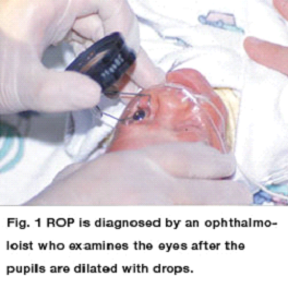
Ophthalmologists (Eye MD’s) who are skilled in the evaluation of infant eyes make the diagnosis of ROP. They examine the eyes after the pupils are dilated with drops. There is active research which is evaluating the effectiveness of digital photography for diagnosing ROP. Infants less than 1500 grams (3.3 lbs) and with a gestational age less than 31 weeks undergo eye examinations to monitor for ROP [See figure 1].

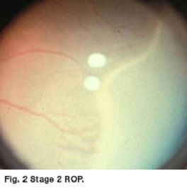
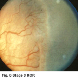
ROP is described by its location in the eye (the zone), by the severity of the disease (the stage) and by the appearance of the retinal vessels (plus disease). The first stage of ROP is a demarcation line that separates normal from premature retina. Stage 2 is a ridge which had height and width. Stage 3 is growth of fragile new abnormal blood vessels. As ROP progresses the blood vessels may engorge and become tortuous (plus disease) [See figures 2 and 3].
When ROP reaches a certain level of severity, called type 1, the potential for retinal detachment (and possible permanent vision loss) becomes great enough to warrant consideration of laser treatment. Eyes that develop this disease have type 1 ROP and are usually treated.

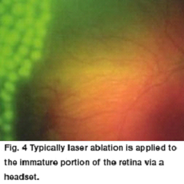
Typically, laser ablation is applied to the immature portion of the retina via a headset [See figure 4].
The outcome of laser treatment is usually favorable with disappearance of the abnormal blood vessels and resolution of plus disease. Despite accurate diagnosis and timely laser treatment, the ROP sometimes continues to worsen and the retina pulls away from the back of the eye. Eyes with retinal detachment caused by ROP generally have a poor visual prognosis. Retinal detachment can be treated with vitrectomy and/or scleral buckling procedure. There is active research in the use of medications to retard the growth of the abnormal blood vessels. These medications may be used as an alternative to, or in addition to, laser treatment. Further research is needed to help determine when and in whom these medications should be used. Despite optimal treatment, some eyes with ROP progress to permanent and severe vision loss.
It is VERY IMPORTANT to have eye exams after discharge from the hospital since ROP may not be resolved before discharge. Also, even with successful treatment of ROP, prematurity may lead to other vision abnormalities. Prematurity is a risk factor for the development ofamblyopia (lazy eye), eye misalignment (strabismus), the need for glasses (even at a young age), and cortical visual impairment. Therefore, every premature infant needs the long-term attention of an ophthalmologist (Eye MD).
Reference: American Association for Pediatric Ophthalmology and Strabismus

